42 dna labeling diagram
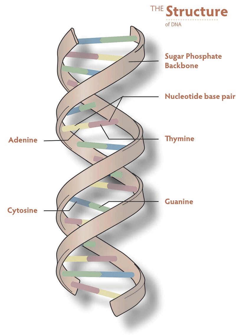
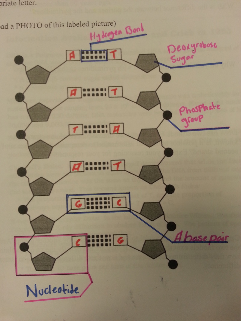
5.2: Structure and Replication of DNA - Biology LibreTexts DNA Structure Watson and Crick proposed that DNA is made up of two strands that are twisted around each other to form a right-handed helix. The two DNA strands are antiparallel, such that the 3ʹ end of one strand faces the 5ʹ end of the other (Figure 5.2. 6 ). The DNA Structure Model - A History of the Double Helix DNA molecules have two strands that coil around an axis, forming a twisted ladder shape, commonly known as a double helix. The rungs on the ladder are the nitrogenous base pairs connected by a hydrogen bond (bases adenine and thymine or guanine and cytosine) while the sides are alternating sugar molecules and phosphate groups bonded together.
Non-invasive DNA-labeling tool opens doors for new research Dutch researchers have developed a new tool to label DNA for studying chromosomes in live cells. The tool is non-invasive and can be applied in culture but also in living organisms, such as ...
Dna labeling diagram
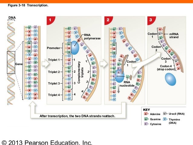
Label-Free Detection of DNA Supramolecular Structure Formation by ... Surface-enhanced Raman spectroscopy (SERS) is widely used to detect the secondary structure of simple DNA molecules, but its application in the revealing of complex DNA supramolecular information remains challenging. Herein, we proposed a modified SERS-based platform able to provide structural information on DNA supramolecular materials. DNA Template Strand | Coding Strand vs. Template Strand - Study.com First, it is important to clarify the structure of DNA, and how it is used in the cell. DNA is double-stranded molecule. Each strand carries all the information needed to create every protein the... 14.2: DNA Structure and Sequencing - Biology LibreTexts The important components of the nucleotide are a nitrogenous base, deoxyribose (5-carbon sugar), and a phosphate group (Figure 14.2. 1 ). The nucleotide is named depending on the nitrogenous base. The nitrogenous base can be a purine such as adenine (A) and guanine (G), or a pyrimidine such as cytosine (C) and thymine (T). Figure 14.2. 1
Dna labeling diagram. Deoxyribonucleic Acid (DNA) - Genome.gov DNA is made of two linked strands that wind around each other to resemble a twisted ladder — a shape known as a double helix. Each strand has a backbone made of alternating sugar (deoxyribose) and phosphate groups. Attached to each sugar is one of four bases: adenine (A), cytosine (C), guanine (G) or thymine (T). en.wikipedia.org › wiki › DNA_nanotechnologyDNA nanotechnology - Wikipedia Properties of nucleic acids. Nanotechnology is often defined as the study of materials and devices with features on a scale below 100 nanometers.DNA nanotechnology, specifically, is an example of bottom-up molecular self-assembly, in which molecular components spontaneously organize into stable structures; the particular form of these structures is induced by the physical and chemical ... What Is DNA? Summary, Structure, and Importance - Healthline The two strands of DNA form a 3-D structure called a double helix. When illustrated, DNA looks like a spiral ladder in which the base pairs are the rungs, and the sugar-phosphate backbones are the ... brd.nci.nih.gov › brd › sopLabeling SOP - National Institutes of Health Labeling SOP Page 4 of 22 Size: 1” W x 1” H BSI Report Generic_Lab_Label_Report / Standard DNA Label Report BradySoft Label Name Label: Lab/Standard Labels/ Standard 2D DNA Narrow Sample Label 1x1.lab Describer: DSC/Standard Labels/ Standardized DNA Label.dsc 4.5 Standard 2D Short 3 Across Label (.5” x 1” Label) 1.
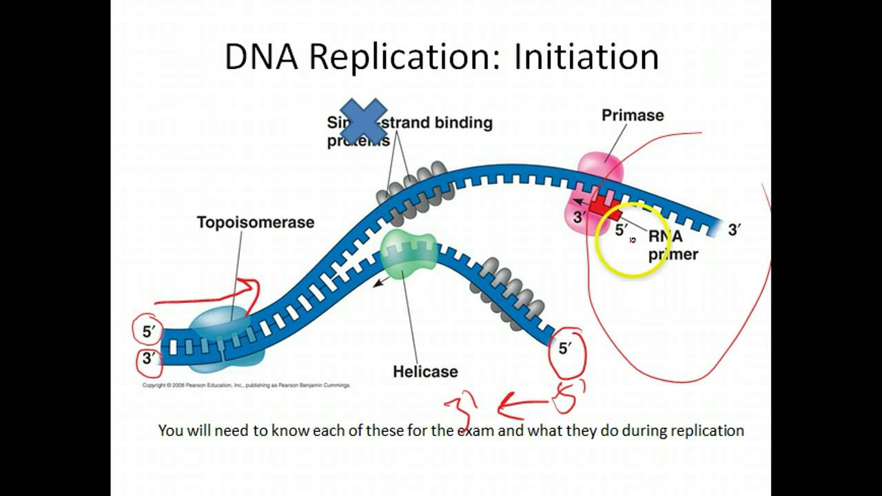
Cisco DNA Center User Guide, Release 2.2.2 Step 1: In the Cisco DNA Center GUI, click the Menu icon and choose Tools > Topology.. Step 2: In the left tree view menu, select the area, site, building, or floor that you are interested in. Step 3: Use the Toggle button to switch between the Geographical map view and the Layer 2 map view.. The Geographical map view displays the sites. DNA Structure - McGraw Hill Education On June 30, 2022, glencoe.mheducation.com and all of its associated sites will be retired and these sites will no longer be accessible. If you wish to retrieve any of the free resources available on glencoe.mheducation.com, please do so prior to June 30, 2022. Label The Diagram Showing Dna Replication. Use These Choices : Bioexcel ... Label the diagrams of dna nucleotides and bases. Dna strand as the dna unwinds. The synthesis of proteins, the central dogma of biology. Label the diagram showing dna replication. Dna ligase dna polymerase leading strand. Dna ligase dna polymerase leading strand 10. In your textbook, read about what dna is and the replication of dna. 10+ Types of Diagrams & How to Choose the Right One - Venngage CREATE THIS TEMPLATE . For more matrix and quadrant chart examples, visit our post on the 20+ SWOT templates, examples and best practices. Return to Types of Diagrams list . Venn diagram. Venn diagrams look like two or more overlapping circles, with text in each section of each circle that describes the categories. With these diagrams, you can quickly communicate differences and similarities ...
› difference › Transcription_vsTranscription vs Translation - Difference and Comparison | Diffen For Transcription, RT-PCR, DNA microarray, In-situ hybridization, Northern blot, RNA-Seq is quite often used for measurement and detection. For Translation, western blotting, immunoblotting, enzyme assay, Protein sequencing, Metabolic labeling, proteomics is used for measurement and detection. Label The Diagram Showing The Dna Replication Use The Choices ... Label the diagram showing the dna replication. This labeled the parental dna. Show how the new strands would be synthesized, using rectangles to represent rna primers and arrows to represent new dna being made. Roles of dna polymerase, primase, ligase, helicase and. Figure 9.9 the semiconservative model of dna replication is shown. Interphase- Definition, Stages, Cell cycle, Diagram, Video G1 and G2 phase represents the time of growth and preparation for mitosis. The synthesis (S) phase is the phase of cell copying or cell duplication of its DNA of its entire genome. Gap 1 (G1) This is the phase in which the cell undergoes normal growth and cell function synthesizing high amounts of proteins. Rna Labelled Diagram - dna sequencing bioninja, rna s role, recent ... all about dna 2 rna structure. Rna Labelled Diagram. Here are a number of highest rated Rna Labelled Diagram pictures on internet. We identified it from reliable source. Its submitted by management in the best field. We assume this kind of Rna Labelled Diagram graphic could possibly be the most trending subject later than we portion it in ...
Double Helix: The Supporting Cast – LSF Magazine – Medium
Nucleotide - Genome.gov A nucleotide is the basic building block of nucleic acids (RNA and DNA). A nucleotide consists of a sugar molecule (either ribose in RNA or deoxyribose in DNA) attached to a phosphate group and a nitrogen-containing base. The bases used in DNA are adenine (A), cytosine (C), guanine (G) and thymine (T). In RNA, the base uracil (U) takes the ...
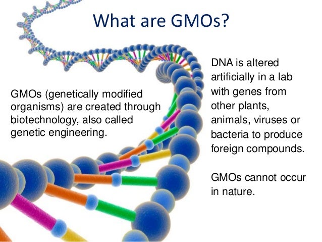
GMO Food and I-522 Labeling
Parts of the brain: Learn with diagrams and quizzes - Kenhub Labeled brain diagram. First up, have a look at the labeled brain structures on the image below. Try to memorize the name and location of each structure, then proceed to test yourself with the blank brain diagram provided below. Labeled diagram showing the main parts of the brain.

DNA Replication Fork. - YouTube
Label The Diagram Showing Dna Replication - Blogger Label the diagram showing the dna replication. Show dna replication with the help of a diagram only. (a) draw a labelled diagram of a \replicating fork\ showing the polarity. Rna primers (label 3′ and. You should label all the parts of the dna including the covalent and hydrogen bonds.
AP Biology: DNA Replication - YouTube
Multiple Choice Quizzes Free Online Biology practice tests Multiple Choice Quizzes, Biology interactive quizzes, AP biology practice test, biology worksheet and labeling quiz

Bacterial Cell Diagrams
Visualizing the invisible: New fluorescent DNA label reveals nanoscopic ... Researchers have developed a new fluorescent label that gives a clearer picture of how DNA architecture is disrupted in cancer cells. The findings could improve cancer diagnoses for patients and ...
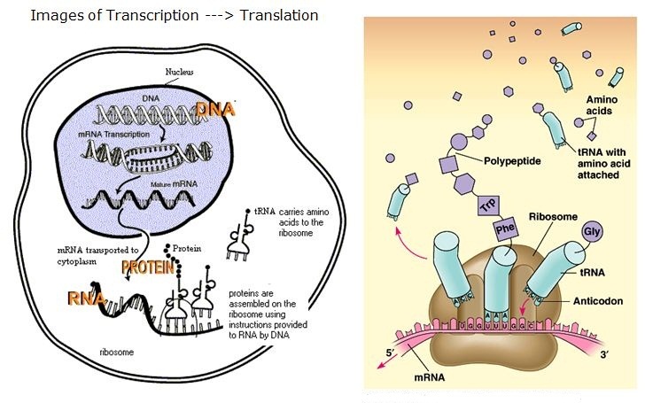
Transcription And Translation Diagram Labeled - Atkinsjewelry
Chromosome dna labeled stock photos and images (186) Educational and medical scheme with cell, chromosome and DNA. Labeled anatomical diagram with cytosine, thymine, adenine and guanine. Stock Photo by normaals 2 / 83 special medical background with a picture of the DNA molecule Pictures by place4design 7 / 307 Vector dna molecule icon Pictures by blumer 2 / 27 DNA strand background.
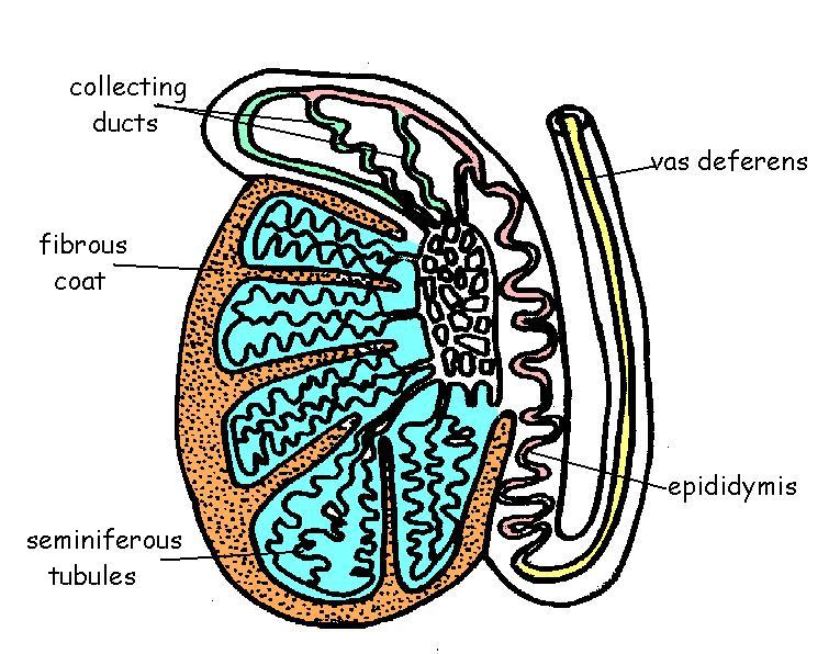
The Anatomy and Physiology of Animals/Reproductive System Worksheet ...
Biochemistry, DNA Replication - StatPearls - NCBI Bookshelf The existence of cell division implies that there is a mechanism that replicates DNA and supplies identical copies for the daughter cells while still maintaining an accurate representation of the genome. This mechanism, known as DNA replication, occurs in all organisms and allows for genetic inheritance. It can occur in a short period, copying up to approximately ten to the 11th power (10^11 ...
DNA - Principle of Biomedical Science
Cladogram- definition, features, parts, examples (vs Phylogram) Cladogram Definition. A cladogram is the graphical representation of the hypothetical relationship (phylogenetic relationship) between different groups of organisms. It is used in the phylogenetic analysis of organisms to determine the evolutionary relationship between them. The cladogram is derived from Greek words clados and gramma where ...
163 ch 03_lecture_presentation
How you can Label a DNA Structure - Biology | ScienceBriefss.com For example, one nitrogenous base of adenine should be labeled Adenine (A), and the attached nitrogenous base of thymine should be labeled Thymine (T). If these labels are clearly presented, the rest of the adenine bases may be marked with an A label and the partner thymine can be marked with T.
Post a Comment for "42 dna labeling diagram"