40 ear diagram
The Ear - TeachMeAnatomy The Middle Ear. View Article. The Inner Ear. View Article. Anatomy Video Lectures. START NOW FOR FREE. TeachMe Anatomy. Part of the TeachMe Series. The medical information on this site is provided as an information resource only, and is not to be used or relied on for any diagnostic or treatment purposes. This information is intended for ... Ear Canal Diagram, Pictures & Anatomy | Body Maps External acoustic meatus. The ear canal, also called the external acoustic meatus, is a passage comprised of bone and skin leading to the eardrum. The ear is comprised of the ear canal (also known ...
Ear Anatomy, Diagram & Pictures | Body Maps - Healthline Inner ear: The inner ear, also called the labyrinth, operates the body's sense of balance and contains the hearing organ. A bony casing houses a complex system of membranous cells. The inner ear ...

Ear diagram
Ear anatomy: Parts and functions - Kenhub The ear is a complex part of an even more complex sensory system. It is situated bilaterally on the human skull, at the same level as the nose. The main functions of the ear are, of course, hearing, as well as constantly maintaining balance. The ear is anatomically divided into three portions: External ear. Middle ear. Blank ear diagrams and quizzes: The fastest way to learn - Kenhub Take a moment to look at the ear model labeled above. This shows you all of the structures you've just learned about in the video, labeled on one diagram. Seeing them all together in this way is a great way to learn, since anatomical structures do not exist in isolation. That's why labeling the ear is an effective way to begin your revision. How to Draw Human Ear Diagram With Labelling #HumanEar Thanks for watching our Channel. how to draw internal structure of human ear,diagram of human ear for class 8,diagram of human ear with labelling,structure o...
Ear diagram. Ear Anatomy: Understanding the Outer, Middle, and Inner Parts of the Ear Tympanic Membrane or Eardrum. The tympanic membrane, or eardrum is the final hearing organ in the outer ear, separating it from the middle ear. The eardrum collects sound waves and vibrates, passing the sound waves into the middle ear. Most hearing disabilities are caused by trauma or disorders in the tympanic membrane eardrum. Label Parts of the Human Ear - University of Dayton Label Parts of the Human Ear. Select One Auditory Canal Cochlea Cochlear Nerve Eustachian Tube Incus Malleus Oval Window Pinna Round Window Semicircular Canals Stapes Tympanic Membrane Vestibular Nerve. Select One Auditory Canal Cochlea Cochlear Nerve Eustachian Tube Incus Malleus Oval Window Pinna Round Window Semicircular Canals Stapes ... Human Ear Anatomy - Parts of Ear Structure, Diagram and Ear Problems Human ear. The ear is divided into three anatomical regions: the external ear, the middle ear, and the internal ear (Figure 2). The external ear is the visible portion of the ear, and it collects and directs sound waves to the eardrum. The middle ear is a chamber located within the petrous portion of the temporal bone. Picture of the Ear: Ear Conditions and Treatments - WebMD Earache: Pain in the ear can have many causes. Some of these are serious, some are not serious. Otitis media (middle ear inflammation): Inflammation or infection of the middle ear (behind the ...
Ear Diagram | Etsy Check out our ear diagram selection for the very best in unique or custom, handmade pieces from our prints shops. Human Ear Diagram - Bodytomy The Structure of Human Ear. Helix: It is the prominent outer rim of the external ear. Antihelix: It is the cartilage curve that is situated parallel to the helix. Crus of the Helix: It is the landmark of the outer ear, situated right above the pointy protrusion known as the tragus. Auditory Ossicles: The three small bones in the middle ear ... Ear pathology Notes: Diagrams & Illustrations | Osmosis This Osmosis High-Yield Note provides an overview of Ear pathology essentials. All Osmosis Notes are clearly laid-out and contain striking images, tables, and diagrams to help visual learners understand complex topics quickly and efficiently. Find more information about Ear pathology: Eustachian tube dysfunction. Otitis externa. The Normal Ear - Understanding Parts of the Ear and How We Hear Outer ear; Middle ear; Inner ear; View the diagrams below to learn more about the different sections of the ear and how we hear. Parts of the Outer Ear The outer ear consists of the visible portion on the side of the head, known as the pinna [1], and the external auditory canal (ear canal) [2].
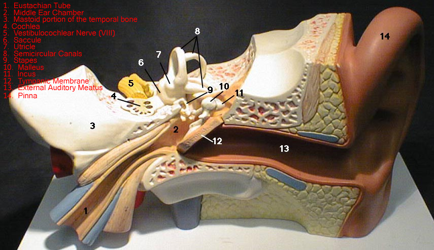
PDF the diagram - Central Institute for the Deaf the diagram EAR HOW WE HEAR 1. Sound enters the ear. 2. The ear drum vibrates. 3. The bones in the middle ear move. 4. The fluid inside the cochlea moves. 5. The hair cells inside the cochlea vibrate. 6. The auditory nerve is activated. 7. The message is sent to the brain. Outer Ear Middle Ear Inner Ear Ear Diagram - Concha Audiology The cochlea is a fluid-filled organ essential for the transduction of mechanical (vibration) energy to electrical (nerve impulse) energy. Vibrations from the stapes on the oval window cause waves within the fluid, which causes the basilar membrane to move. The movement of the basilar membrane causes a shearing action of hair cells (outer and ... human ear | Structure, Function, & Parts | Britannica human ear, organ of hearing and equilibrium that detects and analyzes sound by transduction (or the conversion of sound waves into electrochemical impulses) and maintains the sense of balance (equilibrium). The human ear, like that of other mammals, contains sense organs that serve two quite different functions: that of hearing and that of postural equilibrium and coordination of head and eye ... Anatomy of the Ear | Inner Ear | Middle Ear | Outer Ear The middle ear includes: eardrum. cavity (also called the tympanic cavity) ossicles (3 tiny bones that are attached) malleus (or hammer) - long handle attached to the eardrum. incus (or anvil) - the bridge bone between the malleus and the stapes. stapes (or stirrup) - the footplate; the smallest bone in the body.
ear notching - YouTube
The Human Ear - Structure, Functions and its Parts - BYJUS There are three ear ossicles in the human ear: Malleus: A hammer-shaped part that is attached to the tympanic membrane through the handle and incus through the head. It is the largest ear ossicle. Incus: An anvil-shaped ear ossicle connected with the stapes. Stapes: It is the smallest ossicle and also the smallest bone in the human body.

Ear Piercing – Healing, Procedure, Celebrities, Pictures and diagram
Ear Anatomy - Outer Ear | McGovern Medical School Ear Anatomy - Outer Ear. The outer ear comes in all types of shapes and sizes. This structure helps to give each of us our unique appearance. The medical term for the outer ear is the auricle or pinna. The outer ear is made up of cartilage and skin. There are three different parts to the outer ear; the tragus, helix and the lobule. EAR CANAL

Slideshow: Top Problems in Your Mouth
Ear Diagram (English} | CID Free Download An illustrative diagram to use with caregivers and colleagues. CID School; My Account; Contact; 0 Items. About; Resources; Trainings; Consultations; Blog; FAQs; COVID-19; Select Page $ 0.00. Add to cart. Ear Diagram. An illustrative diagram to use with caregivers and colleagues. You may also like… Ear Diagram (Spanish) $ 0.00; The Ling Six ...
Eye and Ear Models
Ear: Anatomy, Human Ear Diagram, Functions, Parts - Embibe Ear: The human ear is the organ that is responsible for hearing and maintaining a sense of balance. Learn about Ear, Parts, Functions, Diagram.

human ears drawing - Google Search | Ear anatomy, Human ear anatomy ...
Ear Diagram Vector Art, Icons, and Graphics for Free Download Browse 38 incredible Ear Diagram vectors, icons, clipart graphics, and backgrounds for royalty-free download from the creative contributors at Vecteezy!
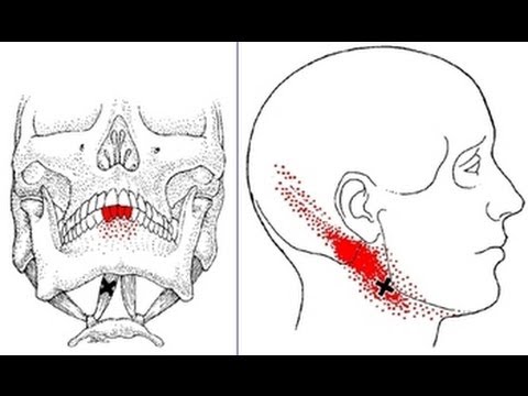
Jaw, Neck, Head, and Teeth Pain from Digastric Muscle Trigger Points ...
The ear canal: Anatomy, diagram, and common conditions The ear canal, or auditory canal, is a tube that runs from the outer ear to the eardrum. The ear has outer, middle, and inner portions. The ear canal and outer cartilage of the ear make up the ...

Origami cat step by step instructions | Page 3 of 5
Human Ear: Structure and Functions (With Diagram) ADVERTISEMENTS: In this article we will discuss about the structure and functions of human ear. Structure of Ear: Each ear consists of three portions: (i) External ear, ADVERTISEMENTS: (ii) Middle ear and (iii) Internal ear. 1. External Ear: It comprises a pinna, external auditory meatus (canal) & tympanic membrane. (i) Pinna: ADVERTISEMENTS: The pinna is […]
Post a Comment for "40 ear diagram"