38 label the diagram of the kidney and nephron below
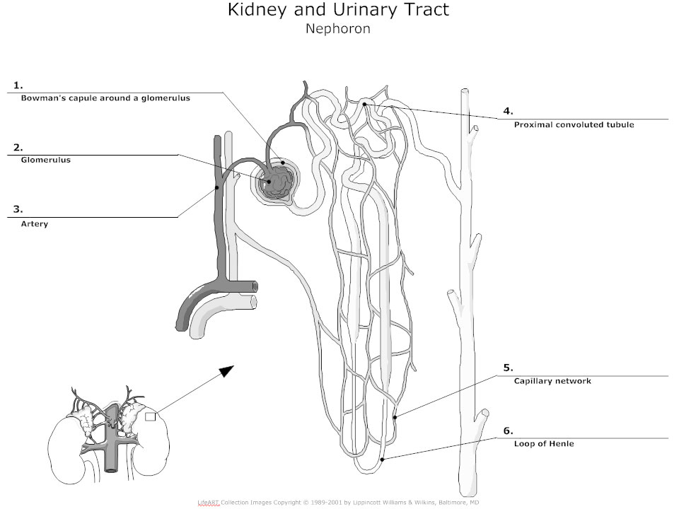
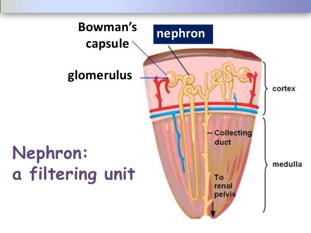
Kidney histology: Nephron, loop of Henle, functions | Kenhub Kidney histology. The kidneys are paired retroperitoneal organs of the urinary system. Their function is to filter blood and produce urine. Each kidney consists of a cortex, medulla and calyces. The nephron is the main functional unit of the kidney, in charge of removing metabolic waste and excess water from the blood. Match Each Lettered Structure In The Diagram Of The Nephron Label these structures in Figure 40C-1 by matching the lettered descriptions with the diagram. a Ureter Carries urine from the kidney. b Renal Each nephron consists of a glomerulus and Bowman's capsule, proximal and distal tubules, the . Figure is a diagram of the nephron and associated blood supply.
25.2 Microscopic Anatomy of the Kidney: Anatomy of the Nephron From old 25.1: Nephrons are the "functional units" of the kidney; they cleanse the blood of toxins and balance the constituents of the circulation to homeostatic set points through the processes of filtration, reabsorption, and secretion. The nephrons also function to control blood pressure (via production of renin), red blood cell production (via the hormone erythropoetin), and calcium ...

Label the diagram of the kidney and nephron below
Diagnosis, Treatment, and Prevention of Hemodialysis Emergencies 7.2.2017 · Most microbubbles <50 μm in diameter and many microbubbles between 50 and 200 μm pass through the venous bubble catcher without triggering an alarm ().The rate of microbubble formation is dependent on the blood flow rate and negative arterial pressure. The total volume of microbubbles during an HD session is a few milliliters, a volume insufficient to cause acute … Blood Filtration in the Kidney - Innerbody 3.7.2018 · The kidneys filter about one-quarter (750-1000 pints) of the blood that is output by the heart daily. This blood is sent to the body’s filter treatment plant, where it is purified by the kidneys and circulated on to the rest of the body. Label the diagram of the kidney and nephron below. - ForNoob Hormones such as antidiuretic hormone (ADH), aldosterone, and atrial natriuretic peptide (ANP) regulate kidney function. Part A - Identifying the structures of the kidney Label the diagram of the kidney and nephron below. Drag the labels to their appropriate locations on the diagram below. Labels can be used once, more than once, or not at all.
Label the diagram of the kidney and nephron below. Pearson Edexcel International GCSE Tuesday 8 January 2019 9.1.2019 · Please check the examination details below before entering your candidate information Other names. 2 ... The diagram shows a human sperm cell. (i) Label the nucleus of this sperm cell. (1) ... Describe the structure of the kidney. Refer to … Life Processes Class 10 Important Questions and Answers 3.8.2020 · (a) Draw a diagram of an excretory unit of human kidney and label the following : Bowman’s capsule, Glomerulus, collecting duct, Renal artery. ‘ (b) Write the important function of structural and functional unit of kidney. (c) Write any one function of an artificial kidney. (CCE 2011) Answer: (a) (b) Nephron. Kidney and Nephron Anatomy Quiz - Registered Nurse RN This is a quiz on the anatomy of the kidney and nephron. Before you start studying the renal system for NCLEX, it is very important you understand the basic anatomy and physiology of the kidney and nephron. These structures are affected by disease processes of the renal system and can lead to various signs and symptoms. This quiz and review will start our renal series. The given diagram represents a nephron. and its bind supply. Study the ... Click here👆to get an answer to your question ️ The given diagram represents a nephron. and its bind supply. Study the diagram and answer the following questions(i) Label parts 1, 2, 3 and 4. (ii) State the reason for the high hydrostatic pressure in the glomerulus. (iii) Name the blood vessel which contains the least amount of urea in this diagram.
Renal Assessment Assignment el.pdf - Kidney/Nephron... - Course Hero Label all structures of the nephron correctly on the diagrams below. A Collecting duct B Descending loopC Ascending loop D Renal vein E Bowmans capsule F Glomerulus G Renal artery H Proximal convoluted tubule I Distal tubule J Capillaries 4. Structure of the Kidney (With Diagram) | Organs | Human Physiology Nephron: Nephron is the basic unit of kidney. The minute structure of the kidney is composed of a number of nephrons. Each human kidney possesses about 1 -2 millions of nephrons. Each nephron is made up of two main parts: (1) Malpighian Body, (2) Renal tubule. (C) Blood Vessels: The two important blood vessels of the kidney are: (1) Renal Artery Part A - Identifying the structures of the kidney Label the diagram of ... Part A - Identifying the structures of the kidneyLabel the diagram of the kidney and nephron below. Drag the labels to their appropriate locations on the diagram below. Labels can be used once, more than once, or not at all. Part B - Water conservation by the kidney diagramstore.com - choose a diagram Diagram of the kidney nephron Diagram of the kidney nephron, what is the kidney nephron structure? The nephron is the. ... Diagram of the human eye and label Diagram of the human eye and label is given below. Cornea The clear. Read more. excel template .
Part A - Identifying the structures of the kidney Label the diagram of ... Label the diagram of the kidney and nephron below. Drag the labels to their appropriate locations on the diagram below. Labels can be used once, more than once, or not at all. Part B - Water conservation by the kidney. The kidneys of terrestrial mammals conserve water in the body by concentrating urine. The osmolarity of human blood is 300 mOsm ... Nephron Drawing Labeled - Labeled Nephron Diagram | Swen Herz Kidney nephrons are the functional units of the kidneys (figure 2). Drawing of a nephron, with labels pointing to glomerulus, tubule, filtered blood, blood with wastes. Bowman's capsule, glomerulus, afferent arteriole, efferent arteriole, renal artery, renal vein, proximal convoluted tubule. The diagram below illustrates the structure of the kidney nephron (a ... The diagram below illustrates the structure of the kidney nephron (a) Name the part labeled E. - Tutorke Get premium membership and access revision papers with marking schemes, video lessons and live classes. Form 2 Biology questions and answers on excretion and homeostasis The diagram below illustrates the structure of the kidney nephron Renal Tubular Acidosis and Management Strategies: A … 26.12.2020 · A schematic diagram illustrating the underlying kidney tubule defects causing the different types of renal tubular acidosis (RTA). Distal (type 1) RTA is caused by either impaired hydrogen (H + ) secretion by vacuolar (v) H + -ATPase or H + /K + -ATPase or increased H + permeability of luminal membrane by α-intercalated cells of the collecting duct, which leads to a …
Kidney Diagram Nephron — UNTPIKAPPS
Biology Questions and Answers Form 2 - High School Biology … It acts on kidney tubules (nephron) ... (glucose) falls below normal. when glucagon. stimulates the liver/when glucagon is produced. after strenuous ... Biology Diagrams to Label - Biology Diagram of Female Reproductive System - Biology Diagrams Pdf - Biology Diagrams in Form 1 - Biology Diagrams in Form 2 - Biology Diagrams in Form 3 ...

32 Label The Diagram Of The Kidney And Nephron Below. - Wiring Diagram ...
Urinary System - Label the Kidney and Nephron Students drag labels to the structures on the slide. Also, the diagram shows the relationship between the aorta, vena cava, and the renal vessels. While these aren't part of the urinary system, they are important in the physiology of the kidney. On the second slide, viewers see a close-up of a kidney that's been cut to show the internal structures.

Nephron Regions | Interactive Worksheet by University of AZ Anatomy and ...
Important Question for Class 10 Science Life Processes - Learn CBSE 16.8.2020 · 12.Draw a neat diagram of excretory system of human beings and label on it: (i) Left kidney (ii) Urinary bladder Answer. 13. Draw a diagram of human respiratory system and label on it : (i) Diaphragm (ii) Larynx Answer. 14.(a) Name the site of exchange of material between the blood and surrounding cells.

31 Kidney Diagram To Label - Labels Database 2020
Solved Kidney Structure and Function The excretory system of - Chegg Hormones such as antidiuretic hormone (ADH), aldosterone, and atrial natriuretic peptide (ANP) regulate kidney function. Part A - Identifying the structures of the kidney Label the diagram of the kidney and nephron below. Drag the labels to their appropriate locations on the diagram below. Labels can be used once, more than once, or not at all.

Kitty Tubes - Digestive System, Reproductive System, and Urinary System ...
Blank Nephron Diagram - schematron.org On the diagram below label the major parts of the kidney. Fill in the blanks below to trace the flow of fluid through a cortical nephron. The first line, . Discover best Nephron Diagram images and ideas on Bing. Updated Nephron Urinary System Diagram; Blank Diagrams Blank Kidney Nephron Diagram.
Urinary System Diagram - Kidney, Urinary Tract, Renal System Diagrams ...
Exam 4 Ch 44 Flashcards - Quizlet Label the diagram of the kidney and nephron below. Drag the labels to their appropriate locations on the diagram below. Labels can be used once, more than once, or not at all. The nephron is the functional unit of the kidney, meaning that filtrate is processed in the nephrons.
Kidneys
Color and Label the Nephron - Pinterest Color and label the nephron, a structure that filters blood within the kidney. Find this Pin and more on Worksheets by Lindsay Sewell. A description of the kidney and how it functions is included with a picture of the kidney and the nephron that students can color. This is a very specific worksheet suitable for advanced biology, anatomy, or ...

explain the structure of nephron with labelled diagram - Biology ...
Nephron - Structure, Functions and Types of Nephron - BYJUS A nephron is the basic structural and functional unit of the kidney. They are the microscopic structure composed of a renal corpuscle and a renal tubule. The word nephron is derived from the Greek word - nephros, meaning kidney. There are about millions of nephrons in each human kidney. Structure of Nephron
Post a Comment for "38 label the diagram of the kidney and nephron below"